Introduction
Fluid inclusions are the original geological fluids trapped within mineral lattice defects and cavities. They serve as valuable records of ancient fluid environments, providing insights into the conditions under which minerals formed, the composition of the fluid, and the sources of materials during different geological periods.
Artificial inclusions, designed to mimic natural ones, have become a powerful tool for understanding various fluid inclusion-related phenomena. These synthetic samples act as standards for instrument calibration and testing methods, and they are increasingly accepted in scientific research. The most mature technique involves creating inclusions by sealing cracks in minerals within an aqueous environment, with the H2O-CO2 (±NaCl) system being the most widely used.
Raman spectroscopy is a non-invasive analytical technique that relies on inelastic scattering caused by molecular and lattice vibrations. It offers advantages such as rich spectral information, high efficiency, low sample consumption, and minimal damage to the sample. Laser micro-Raman spectroscopy combines advanced spectroscopy, chemometrics, detection technology, and computer systems. In the field of fluid inclusions, it provides several benefits: non-destructive and contactless analysis, high sensitivity, detailed spectral data, and the ability to study phase equilibrium, mineral dissolution, and deep rock partial melting in situ.
This paper uses Raman spectroscopy to analyze synthetic fluid inclusions and compares them with natural ones to verify the feasibility of this method in fluid inclusion studies.
Current Status of Inclusion Raman Spectroscopy
Fluid inclusions contain trace amounts of the original ore-forming fluid and form a relatively closed system, making them ideal for studying the original fluid properties. Their formation pressure is crucial for understanding oil and gas migration, accumulation history, and tectonic activity. Several methods are currently used to measure the pressure of fluid inclusions, including the isothermal diagram method, salinity-temperature method, sodium chloride-water solution density method, isovolumetric method, and PVT simulation.
Since the 1970s, Raman spectroscopy has been applied to fluid inclusions. Rosasco et al. (1975) were among the first to publish results on natural inclusions. Beny et al. (1982) and Tourary et al. (1985) further expanded the use of Raman techniques in fluid systems. These studies not only demonstrated the potential of Raman spectroscopy but also laid the foundation for quantitative analysis. Pasteris et al. (1988) discussed the limitations of Raman instruments and optimized analytical conditions, promoting broader application of the technique.
In China, researchers such as Huang Weilin (1990) and Xu Peicang (1996) used Raman spectroscopy for fluid inclusion analysis and explored quantitative methods in detail.
Laser Raman Detection Principle
Raman spectroscopy, with nearly 90 years of development, was theoretically predicted by A. Smekal in 1923 and experimentally discovered by C.V. Raman in 1928. It involves the inelastic scattering of light due to molecular and lattice vibrations. The technique has gained widespread use in fields such as biotechnology, environmental monitoring, medicine, and material science.
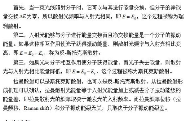
The Raman effect occurs when a laser excites a sample, causing elastic and inelastic scattering. Most molecules undergo elastic collisions, while a small fraction experiences inelastic collisions, resulting in frequency changes. This leads to either Stokes or anti-Stokes scattering, depending on whether energy is lost or gained. The Raman shift depends on the vibrational energy levels of the molecule, not the incident frequency.

Figure 1: Raman and Rayleigh Scattering Energy Levels
Experimental Procedure
Sample preparation involved a fusion silica capillary technique, producing inclusions with diameters of 8–12 mm. To optimize Raman signal quality, samples were ground to a thickness of 50–200 µm and mounted using beeswax. High reflectivity minerals like calcite can cause double imaging, making sample identification difficult. For optimal results, inclusions near the surface (depth 30–70 µm) were preferred to minimize background interference and maximize signal strength. Confocal Raman spectrometers are less affected by these issues, so they were chosen for this study.
Fluorescence from surfaces, matrix minerals, and inclusions can interfere with measurements, so test conditions were carefully selected to avoid such effects.
Equipment used included the Finder Vista Microscopic Confocal Raman Spectrometer System, featuring a high-performance CCD backscatter detector. The system operated at a laser wavelength of 532 nm (10 mW), with a 1800 g/mm grating, resolution <0.9 cmâ»Â¹, slit width of 100 µm, integration time of 20 seconds, and a 10× objective lens.
Experimental Analysis
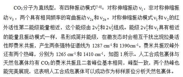
The Raman spectra of natural and synthetic inclusions are shown in Figure 2. The experimental sample contained COâ‚‚ gas inclusions, which provide critical information about mineralization, hydrocarbon migration, fluid evolution, and tectonic dynamics.
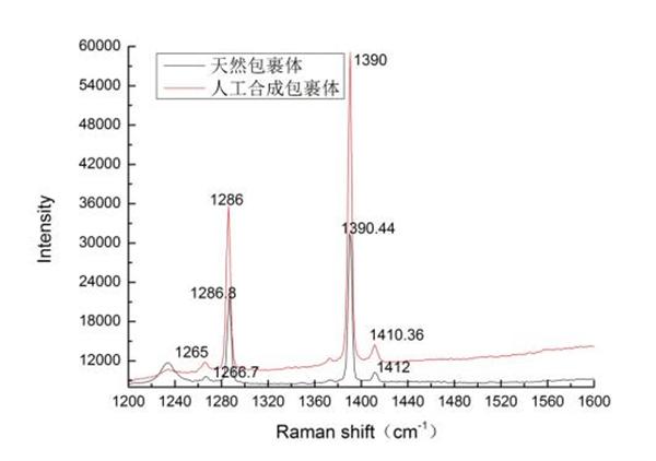
Figure 2: Raman Spectra of Natural and Synthetic Inclusions
Conclusion
Raman spectroscopy offers a simple, direct, fast, and precise method for analyzing capillary samples. It does not interfere with fluid signals and allows accurate focusing on each phase, ensuring high-quality Raman signals. Synthetic inclusions provide clear insights into phase changes, supporting accurate observations of natural inclusions. COâ‚‚ synthetic inclusions serve as standard samples for verification and analysis, offering technical feasibility and practicality for Raman spectroscopy of natural fluid inclusions.
References
[1] Li Jiajia, Li Rongxi, Liu Haiqing. Research progress in the determination of fluid inclusion pressure by laser Raman spectroscopy [J]. Physical and Chemical Testing - Chemistry, 2016, 52(7): 859-864.
[3] Ni Pei, Ding Junying, I-Ming Chou et al. A new type of artificial "fluid inclusion": fusion silica capillary technology [J]. Geoscience Frontiers, 2011, 18(5): 132-139.
[4] Chen Yong, ERNSTA A. J. Burke. Principles, methods, existing problems and future research directions of laser inclusion Raman spectroscopy for fluid inclusions [J]. Geological Review, 2009, 55(6): 851-861.
[5] Chen Jinyang, Zheng Haifei, Zeng Yushan et al. In situ analysis of fluids at high temperatures using inclusions as cavities[J]. SPECTROSCOPY AND SPECTRAL ANALYSIS, 2003, 23(4): 726-729.
[6] Chen Yong. Principles, methods, existing problems and future research directions of laser inclusion Raman spectroscopy for fluid inclusions [J]. Geological Review, 2009, 55(6): 851-860.
[7] Ding Junying, Ni Pei, Guan Shenjin. Hâ‚‚O-COâ‚‚ In-situ Micro-laser Raman Spectroscopy Study of System Fusion Silica Capillary Samples[J]. Geoscience, 2011, 18(5): 140-146.
[8] Li Jiajia, Li Rongxi, understand, etc. Microscopic laser Raman quantitative analysis of COâ‚‚ Gas Carbon Isotope Composition Method[J]. SPECTROSCOPY AND SPECTRAL ANALYSIS, 2016, 36(8): 2391-2398.
48V Battery Pack ,Switch Battery Pack,Portable Lithium Battery Pack,48V Lithium Ion Battery Pack
Zhejiang Casnovo Materials Co., Ltd. , https://www.casnovonewenergy.com